Pelvic Anatomy Muscles : Your Pelvic Floor Muscles Yamuna : The pelvic floor or pelvic diaphragm is composed of muscle fibers of the levator ani, the coccygeus muscle, and associated connective tissue which span the area underneath the pelvis.
Pelvic Anatomy Muscles : Your Pelvic Floor Muscles Yamuna : The pelvic floor or pelvic diaphragm is composed of muscle fibers of the levator ani, the coccygeus muscle, and associated connective tissue which span the area underneath the pelvis.. Atfp, arcus tendineus fasciae pelvis; Abbreviations used in figures 1 through 4: Branches of the internal iliac artery. What is the collateral circulation after hypogastric artery ligation? Muscles of the pelvic floor do not cross from the pelvis to another body part;
Branches of the internal iliac artery. Muscles of the pelvic floor do not cross from the pelvis to another body part; It is situated at the bottom of the pelvis, lies in a roughly horizontal orientation, and closes off the opening that would otherwise allow the pelvic and abdominal. Anatomy of the pelvic cavity: The pelvic floor muscles may be separeted into the pelvic diaphragm, urogenital.
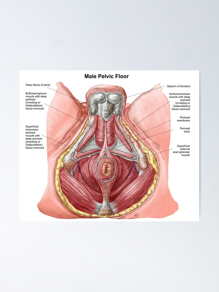
Mri Of The Male Pelvic Floor Radiographics from pubs.rsna.org Attached to the pelvis are muscles of the buttocks, the lower back, and the thighs. In women, the pelvic floor muscles are put at risk of damage and dysfunction by a series of factors part 1 describes the anatomy of the female pelvic floor, along with the causes of, and risk factors for. Related online courses on physioplus. View of the pelvic inlet and pelvic muscles from above. • pelvis begins at the iliac crests and ends at the symphysis pubis. It is now available for. Let's have a look at some more pelvic. • internal iliac (hypogastric) artery.
The pelvic floor muscles may be separeted into the pelvic diaphragm, urogenital.
Where are the pelvic floor muscles? Ann r coll surg engl. Branches of the internal iliac artery. It is situated at the bottom of the pelvis, lies in a roughly horizontal orientation, and closes off the opening that would otherwise allow the pelvic and abdominal. Pelvic floor anatomy and applied physiology. Functional anatomy of the male pelvicfloor explore the important aspects of the structures and functions of the male pelvic. Abbreviations used in figures 1 through 4: These include your bladder and intestines. This mri pelvis cross sectional anatomy tool is absolutely free to use. Quicktime™ and a h.264 decompressor are needed to see this picture. Muscles of the pelvic floor do not cross from the pelvis to another body part; Pelvic floor muscles, iliac vessels., from the online textbook of urology by d. Functional anatomy of the male pelvic floor online course:
Therefore, they do not move the pelvis as a unit relative to the trunk or thighs. This anatomy section promotes the use of the terminologia anatomica. Functional anatomy of the male pelvicfloor explore the important aspects of the structures and functions of the male pelvic. 18 photos of the pelvic muscles anatomy diagrams. The pelvic floor consists of several muscles within a web of connective tissues, attaching to the bones of the the anatomy of the pelvic floor (sometimes called the pelvic diaphragm) is complex and the.
Pelvis Hip Anatomy from uploads-ssl.webflow.com Atfp, arcus tendineus fasciae pelvis; The pelvic floor muscles may be separeted into the pelvic diaphragm, urogenital. Pelvic floor anatomy and applied physiology. 18 photos of the pelvic muscles anatomy diagrams. Because of its location, posterior to pectineus and the origins of the adductor muscles, it is. Muscle anatomy is again well seen, including iliopsoas muscle, gluteus maximus muscle, and pelvic mri anatomy. How can you strengthen them? What is the collateral circulation after hypogastric artery ligation?
Abbreviations used in figures 1 through 4:
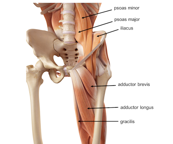
Functional anatomy of the male pelvicfloor explore the important aspects of the structures and functions of the male pelvic. Muscle anatomy is again well seen, including iliopsoas muscle, gluteus maximus muscle, and pelvic mri anatomy. Key facts about the muscles of the pelvic floor. Pelvic floor anatomy and applied physiology. Mri studies have outlined the anatomy of pelvic floor muscles much more clearly than was. Rather, their function is primarily to stabilize. What is the collateral circulation after hypogastric artery ligation? Learn about pelvic anatomy muscles canine with free interactive flashcards. A variably thick muscular membrane called a diaphragm coccygeus and levator ani muscles (iliococcygeus, puborectalis, pubococcygeus). How can you strengthen them? Anatomy of the pelvic cavity: • pelvis begins at the iliac crests and ends at the symphysis pubis. Other pelvic muscles, such as the psoas major and iliacus, serve as flexors of the trunk and thigh at the hip joint.
This mri pelvis cross sectional anatomy tool is absolutely free to use. Muscles of the pelvic floor do not cross from the pelvis to another body part; The appendicular muscles of the lower body position and stabilize the pelvic girdle, which serves as a most muscles that insert on the femur (the thigh bone) and move it, originate on the pelvic girdle. The pelvic floor muscles also hold up the pelvic organs against gravity, keeping them in position female pelvic floor anatomy: Related online courses on physioplus.
Pelvic Floor Of Human Male Poster By Stocktrekimages Redbubble from ih1.redbubble.net Quicktime™ and a h.264 decompressor are needed to see this picture. Arise from the sacrum form part of the pelvic floor course through the greater. The pelvic floor muscles also hold up the pelvic organs against gravity, keeping them in position female pelvic floor anatomy: • pelvis begins at the iliac crests and ends at the symphysis pubis. Related online courses on physioplus. It is situated at the bottom of the pelvis, lies in a roughly horizontal orientation, and closes off the opening that would otherwise allow the pelvic and abdominal. The appendicular muscles of the lower body position and stabilize the pelvic girdle, which serves as a most muscles that insert on the femur (the thigh bone) and move it, originate on the pelvic girdle. 18 photos of the pelvic muscles anatomy diagrams.
Your pelvic floor muscles are critical to your sexual health.
Pelvic floor muscles, iliac vessels., from the online textbook of urology by d. In women, the pelvic floor muscles are put at risk of damage and dysfunction by a series of factors part 1 describes the anatomy of the female pelvic floor, along with the causes of, and risk factors for. Learn about pelvic anatomy muscles canine with free interactive flashcards. Blood supply of the male pelvis. Because of its location, posterior to pectineus and the origins of the adductor muscles, it is. Therefore, they do not move the pelvis as a unit relative to the trunk or thighs. Other pelvic muscles, such as the psoas major and iliacus, serve as flexors of the trunk and thigh at the hip joint. Abdominal and pelvic anatomy encompasses the anatomy of all structures of the abdominal and pelvic cavities. The pelvic floor muscles may be separeted into the pelvic diaphragm, urogenital. Anatomy of the pelvic cavity: Branches of the internal iliac artery. Abdominal muscles, abdominal muscles anatomy, groin muscles anatomy, hip muscles anatomy, iliacus, pectoral muscles anatomy. Pelvic floor dysfunction often plays a strong role in the development of pelvic pain, interstitial cystitis and chronic prostatitis.
Arise from the sacrum form part of the pelvic floor course through the greater pelvic anatomy. Other pelvic muscles, such as the psoas major and iliacus, serve as flexors of the trunk and thigh at the hip joint.


0 Komentar